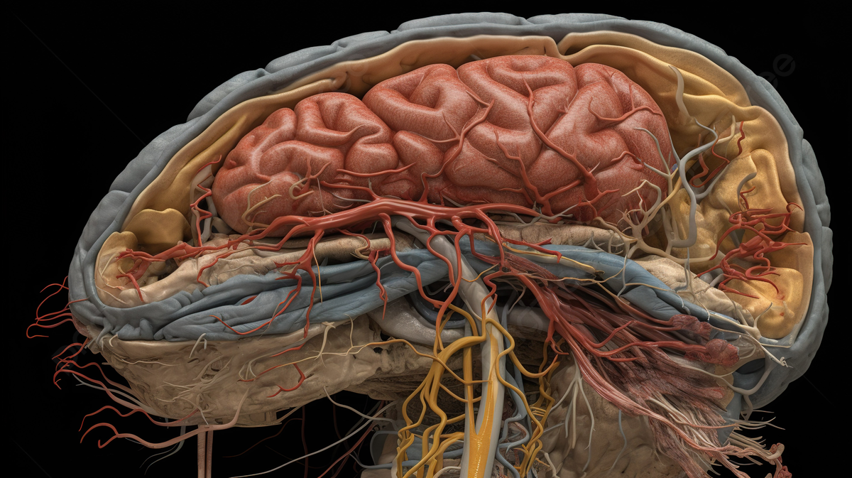
On December 10, researchers from IIT Madras made history by unveiling a 3-D map of five developing fetal brains from the second trimester of pregnancy. These high-resolution 3-D maps provided cellular-level information about the developing human brain. This is the most detailed map of a brain formed between 14 to 24 weeks inside the womb. This is also the time period when the human brain goes through rapid growth in size and complexity.
Project DHARINI
The brain atlas project developed by Sudha Gopalakrishnan Brain Centre at IIT Madras is one of its kind project. Under the name DHARINI, it has captured the developing human brain at a very early stage.
The brain is the most complex organ of our body, and its mapping requires a lot of precision and time. The team of researchers at IIT used advanced technology to visualise approximately 5000 brain slices and more than 500 brain regions, put together. The information gathered by IIT research researchers under project DHARINI is free to access and it will be a valuable resource for neuroscientists across the world.
To top this, precision at a cellular level was achieved while creating the 3-D map of the brain. This will particularly be a significant step forward in understanding brain disorders.
“This is groundbreaking research for clinicians. It will help us study how the human brain develops in the womb. For instance, we have discovered some surprising differences in timelines. What we precisely thought occurred at 14 weeks may actually happen at 17 weeks,” said Dr Kumutha, the dean and professor of neurology at Chennai’s Savitha Medical College and Hospital. She was one of the collaborators on the project.
According to Dr. Kumutha, the data coming out can provide critical insights into brain disorders such as autism. Still not understood very properly, now neuroscientists will be able to understand the development of autism in children as well as adults.
“It may also help explain why some children suffer permanent damage and develop cerebral palsy after hypoxia or lack of oxygen while others recover without lasting effects,” adds Dr Kumutha.
Data coming out from the project will keep Scientists busy for years to come. A better understanding of the human brain will help create precise models for study. In spite of AI being a very important helping hand, precisely collected data from physical experiments will also be required at the moment.
Slicing The Brian
The Sudha Gopalakrishnan Brain Centre inside IIT Madras is supported by Infosys. The company’s co-founder, Kris Gopalakrishnan is trying to better AI tools that can be used to study the brain.
The brain atlas created by researchers of IIT Madras is the largest data set in the world. Alongside, it is the only study that was able to capture a growing brain inside human foetuses.
In 2016, the Allen Institute for Brain Science published a free-to-access brain atlas that captured the brain of an adult woman in approximately 1300 slices. However, the latest study surpasses this 2016 data. Five still borns in the second trimester were used to study and capture the complex structure of the brain.
The brain went through cryogenic freezing and was then thinly sliced for scientists to see and analyse the structure carefully. “The brains were very thinly sliced using complex robotic instrumentation. The slices are of just 10 to 20 microns thickness which is equivalent to 1/10 or 1/5 the thickness of the human hair,” says Professor Mohanshankar Sivaprakasam, head at Sudha Gopalakrishnan Brain Centre.
The thin transparent slices were stained and microscopically imaged in detail. Later, these slices were put together to create a 3-D map. All the instrumentation and technology used for freezing, sizing, creating plates, digiting and mapping were indigenously developed by researchers at IIT Madras.
It took around five years from scratch to finish the complete atlas of the brain by the Allen Institute in 2016. However, the IIT Centre has collected and processed one large brain within a month.


