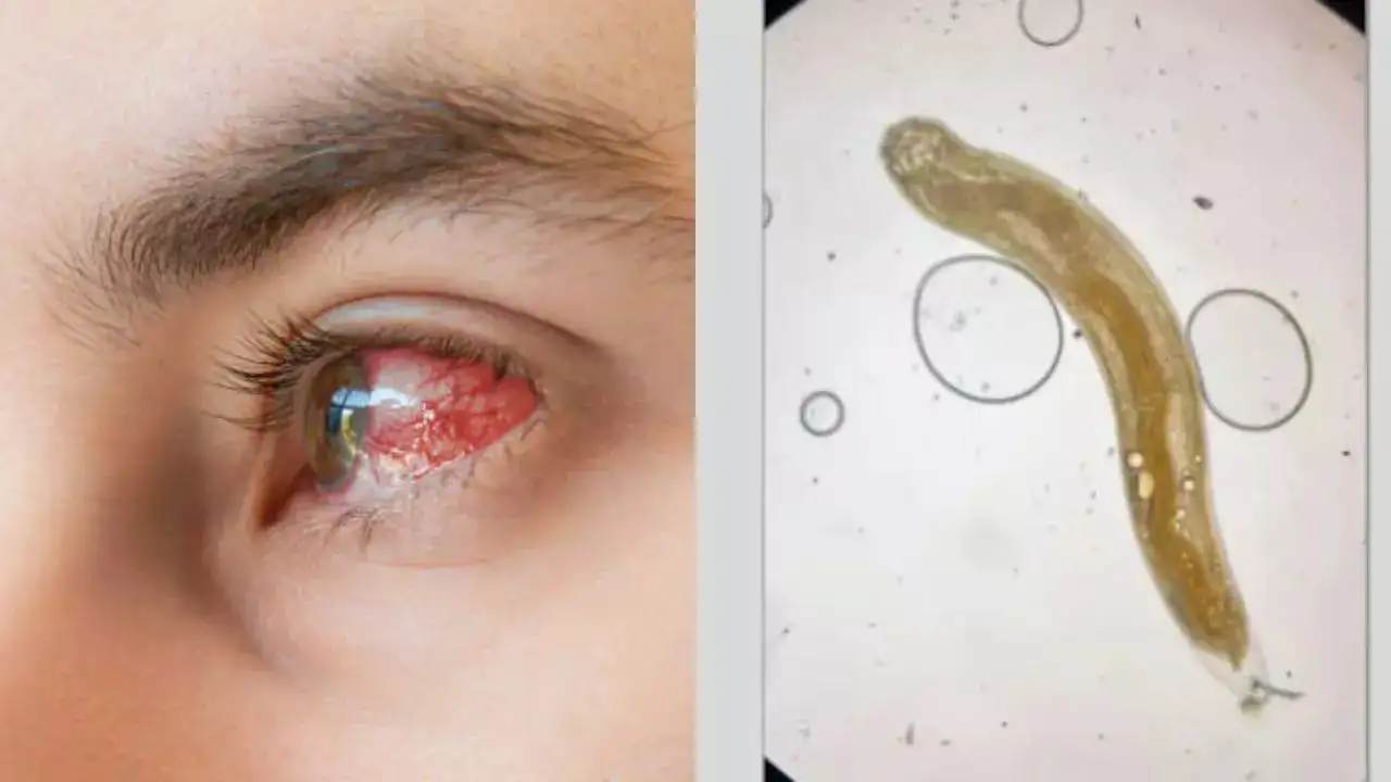
In a shocking incident, doctors pulled a live parasitic worm from a man’s eye in a surgery. The 35-year-old man is from rural central India. He visited an eye clinic after suffering from persistent blurry vision and eye redness for over eight months. What seemed like a common eye infection turned out to be far more serious. The doctors discovered a live, sluggishly moving parasitic worm wriggling in the back of his eye.
A Rare Parasite from an Unlikely Source
Following this the doctors conducted examinations with fundoscopy, ophthalmologists. They found a Gnathostoma spinigerum in the back of the man’s eye. It is a parasitic roundworm typically found in the intestines of cats and dogs.
The man had likely contracted the worm through the consumption of raw or undercooked fish, meat, or poultry. This is a known transmission route in areas where the parasite is endemic.
Complex Eye Surgery conducted
After the discovery, the doctors performed an emergency pars plana vitrectomy (PPV) to remove the parasite. The delicate procedure involved extracting the vitreous humor, the jelly-like substance inside the eye, to reach the worm. According to reports, under a microscope, the parasite was confirmed to be in the larval stage, showing distinctive features like a cephalic bulb, thick outer cuticle, and a well-developed intestine.
The doctors performed pars plana vitrectomy (PPV) to remove the worm. “Under light microscopy, a larval-stage nematode with a cephalic bulb, thick cuticle, and well-developed intestine was identified; these features were consistent with Gnathostoma spinigerum,” the doctors wrote.
Experts comment
“Gnathostomiasis is one of several parasites that can infect the eye and the retina,” Abdhish Bhavsar, MD, a clinical spokesperson for the American Academy of Ophthalmology and a retina specialist at Retina Consultants of Minnesota, told MedPage Today.
“Some of these worms are larger, and some of them are smaller than others, and some of them are very small and can travel within the retina or subretinal space under the retina, and can cause significant damage to the eye and to the vision or even potentially cause blindness.”
Recovery Involved Steroids and Anti-Parasitic Treatment
Post-surgery, the man was treated with oral and ocular steroids to reduce inflammation caused by uveitis (a condition marked by swelling in the eye). He also received a course of albendazole, an anti-parasitic medication. While the infection was cleared, a cataract developed in the affected eye, causing permanent partial vision loss. At an 8-week follow-up, his visual acuity was recorded at 20/40.
What is Ocular Gnathostomiasis?
According to reports, Ocular gnathostomiasis is a rare but serious condition caused by the migration of Gnathostoma larvae into the eye. Although it is uncommon in humans, the infection can lead to severe inflammation, vision damage, and even blindness. Experts warn people in regions like India, Thailand, and Japan to avoid eating undercooked freshwater fish or meat to prevent such infections.


